How to draw female reproductive system


Female reproductive system has muscular elastic bag like structure called Uterus and pair of  ovaries attached to it on either sides. 

Uterus opens into vagina through narrow cervical canal.Two tube like structures emerge out of  uterus on either sides. These are called oviducts.We can start with basic shape of Uterus, later we go for oviducts and vagina. Let`s start   the diagram.

Step 1:
Draw a inverted triangle as shown.


Step 2:
Draw out line of uterus around the triangle as shown.


Step 3:
Erase the inner  triangle as shown.


Step 4:
Draw a smaller triangle  in the middle.


Step 5:
Complete the inner structure of uterus by following
the triangular shape as shown.


Step 6:
Define the tip of uterus as shown to represent cervix.


Step 7:
Draw two tubes  out of uterus on either sides as shown.


Step 8:
Continue the curved line of uterus along the oviducts
as shown.
Draw the vagina from the outer surface of cervix as shown.


Step 9:
Complete the inner lining of vagina with folded pattern


Step 10:
Label the parts neatly as shown.The diagram is complete.
Read more about Human female reproductive system Click here.


How to draw human eye section

Human eye is spherical organ. The front portion is protruding out and it encloses lens, iris. It has three layers namely outer sclera, middle choroid and inner retina. Retina is sensory layer of eye which has millions of  Photo receptors called and Rods and Cones. An optic nerve emerges from lower side of  eye ball.Lens is transparent and is held in position by suspensory ligaments. Entry of light in to eye ball is controlled by Iris which is made up of pigmented smooth muscle.
.Chorioid layer forms transparent cornea before lens.Lets draw the eye ball from a circle. You can make a circle using rounder.

 Step 1:

Draw a  circle and a line through center of circle as shown.
Mark two lines on either sides.
 Step 2:
Erase the portion of  curve.between the two lines.
 Step 3:
Draw an imaginary arch before the eye ball.
Step 4:
Connect the arch on either sides as shown.
 Stop 5:
Clean the extra lines.
 Step 6:
Make a double line along the out line.

 Step 7:

Put a dot to position the lens correctly as shown.


 Step 8:
Draw ciliary muscle on either sides as shown.

 Step 9:
Draw iris as shown.
 Step 10:
Draw suspensory ligaments connecting lens.
 Step 11:
Draw conjunctiva on cornea as shown.

 Step 12:
Draw retina on inner side of the choroid layer.

Step 13:
Erase a little on bottom of eye ball to draw optic nerve.

Step 14:

Draw optic nerve as shown and label the parts neatly.


How to draw chloroplast

Chloroplast is cell organelle which is seen only in plant cells and algal cells. A typical chloroplast is in biconvex shape. It has outer membrane and inner membrane.

The inner membrane encloses viscous fluid called stroma. In stroma there is network of  several piled thylakoids.

Chloroplast is food synthesizing organelle. It prepares food called starch by process called Photosynthesis.

 Step 1:

Draw a double convex shape as shown.


 Step 2:

Draw one more curve upon,  following the contours of below curve.


 Step 3:

Draw about three faint reference  lines inside as shown. These lines help in drawing grana thylakoids in proper orientation.
 Step 4:

Draw about three to five grana along the reference lines as shown.

Step 5:

Connect the grana thylakoids by grana lamellae as shown.

Put several neat dots to represent stroma of chloroplast.


Label the parts neatly as shown.


How to draw Mitochondria

Mitochondria is cell organelle which generates power required for cell and hence called `Power house of the cell". They are sac like structures present in the cytoplasm of the cells. They may be of various shapes like thread, spherical or club Mitochondria have two compartments an inner compartment and an outer compartment.  The viscous substance in the inner compartment is called Matrix. The matrix is surrounded by a membrane called Inner membrane. The inner membrane is thrown into several folds called Cristae. Now let`s draw mitochondria.


 Step 1:
Draw a rough sausage shape with free hand in faint curve. Do not stop the curve in the middle, continue until you meet the starting point.
 Step 2:
To give a sliced sausage look, draw one more curve as shown.

Line inner side with another faint curve as shown.
 Step 3:
Erase the inner line leaving the faint impression and mark the positions of cristae with faint lines.

Observe uniform spacing between the faint lines as shown.
 Step 4:
Draw the inner membrane by following  previous impressions of curve and reference lines of cristae.

Complete it till you meet the other end of the curve.
Step 5:
Tap clean dots with sharp pencil nib to indicate
matrix.

To make diagram much detailed you can also draw mitochondrial DNA and ribosomes.

 Continued :-The cristae extend into matrix. The space between the folds is continuous with the outer compartment. On the inner membrane, projecting into the matrix are a large number of particles called Elementary particles. These particles have a spherical head and a stalk. They are attached to the inner membrane by their stalk and the head portion of the particle  is in matrix.

How to draw TS of leaf


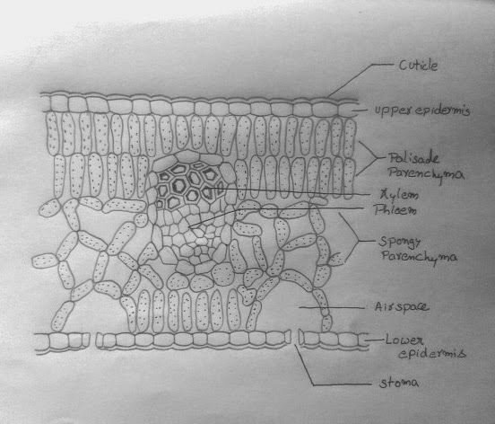
A transverse section of a typical leaf shows that it is covered by epidermis on both surfaces the upper epidermis on the upper surface and lower epidermis on the lower surface. Lower epidermis is interrupted by a large number of opening called stomata. The central opening is called stoma. Each stoma is surrounded by the two kidney-shaped cells called Guard cells. Stomata regulate the exchange  of gases and loss of water vapor from the leaves.Tissue enclosed between the two epidermal layers is called the Mesophyll. Mesophyll consists of elongated cells arranged in rows called the Palisade parenchyma. It also has irregularly arranged cells with large intercellular spaces known as spongy parenchyma. The mesophyll cells contain abundant chloroplasts. More palisade tissue occur in leaves exposed to bright light than those exposed to shade. Vascular bundles in the leaf are located in the midrib and the veins. In each vascular bundle of the leaf, phloem is located towards the lower side and xylem toward the upper side. 

Now let`s start the diagram-
 Step 1:
Draw two faint parallel line as shown.

Draw outlines of central vascular bundle
and air spaces above stomata.

Step2:
Erase the faint lines and draw wavy patter of
epidermis as shown.
 Step3:
Complete the wavy pattern on both sides of  the
diagram as shown. Note that the edges of outer line should face the edge of inner line exactly.

Step 4:
Connect the edges of the lines to create barrel shaped epidermal cells on both sides.
Complete the bundle sheath cells of  vascular bundle too by same technique.
Step 5:
Draw cuticle on both epidermis following the contours of the epidermal cell.
Step 6:
Draw the mesophyll cells lining the air spaces as shown.

Draw palisade tissue as shown along the upper epidermis.
Step 7:
Complete drawing the palisade tissue as shown.
Draw spongy tissue as shown.

Put several dots in all mesophyll cell which represent chloroplasts.

If your leaf specimen has trichomes you can draw on upper epidermis.

 Step 8:
Draw hexagonal cells in vascular bundle in top position as shown. Do not leave any spaces between these cells.

Draw thick cell walls inside xylem with thick lines use little pressure.

Step 9:
Complete phloem as shown with several small hexagonal cells. 

Label the diagram neatly as shown.

Continuation:- Leaf anatomy is suitable for entry of carbon dioxide into the closely packed palisade cells of the leaf. More photosynthetic activity takes place in the upper side of the leaf. On this side, the palisade cells contain more number of chloroplasts than the loosely arranged spongy parenchyma cells towards inner side. Organization of the leaf is suitable for entry of raw materials, synthesis of sugar and transport of the photosynthate. Gaseous carbon dioxide passes from atmosphere into the leaf through diffusion and reaches the chloroplasts. Water is transported from the roots through xylem by veins and its branches called veinlets. There is a large surface area to receive light on leaf lamina. Oxygen produced during photosynthesis diffuses outside through stomata. Sugar is converted into starch in the chloroplast during the day time and starch is to sugar. This sugar will pass through phloem to other parts of the plant.

How to draw a leaf

Leaf is the main photosynthetic organ of plant. It is a laterally, flattened structure borne at a node.It usually has a bud in its axil. Leaf constitutes the main photosynthetic organ of plants owing to the presence of chloroplasts and chlorophyll. A leaf consists to three parts viz Lamina , Petiole and leaf base. The expanded portion of the leaf is called the Lamina. 

Now let`s draw the leaf.

Leaf have four main features which should be considered while drawing. They are petiole, leaf margin, leaf apex and overall shape.Here the leaf
has wavy margin and very small petiole. Observe the reticulated venation and smooth texture of leaf.


Step 1:

Draw oblique mid vein first with free hand.
Roughly draw the margins of the leaf in faint curves as shown.
Represent tiny petiole of leaf also.

Step 2: 

Define the midrib with second line, draw only with free hand.Note that midrib gradually sharpens to tip.

Make the faint lines thick.

Mark the positions of the lateral veins.

 Step 3:

Draw the lateral veins by observing the specimen leaf. Pay close attention to pattern of veins and vein lets on leaf.


 Step 4:

Draw the veinlets as shown and label the parts
clearly.
Continued:-It has a number of veins and veinlets. Prominent vein in  the middle is known as Midrib. Rate of leaf production, thickness of leaf, shape and area of leaves and their orientation to light and periods up to which leaves are retained are some of the factors that determine plant productivity.

How to draw human digestive system


The human digestive system starts with mouth and ends with anus. Intake of food into to mouth is called Ingestion.Mouth encloses a cavity within called Buccal cavity or Oral cavity. Three pairs of salivary glands pour their secretions called saliva in to this cavity. The muscular tongue pushes the food under grinding teeth as it mixes with saliva, this process is called "Mastication". Mastication makes the food into ball of paste. Salivary amylase" an enzyme in saliva starts the digestion of starch right away from mouth. As soft ball of food enters Oesophagus, the wavy movements (Peristaltic movements) drive the bolus into stomach. Oxyntic cells present in inner lining of stomach secrete Hcl acid which has killing action on Microbes and partially denatures the proteins in food.Slimy mucous secreted my mucous glands of stomach protect inner lining from self destruction due to acid. Gastric juices of stomach starts digestion of Proteins, lipids but not carbohydrates.


Human digestive system mainly have three parts to be drawn in proportion they are the Stomach, Liver and Intestines.Students feel difficult while drawing the duodenum, bile duct, Gall bladder and Pancreas in  a narrow space. Even the coiled small intestines look complicated.Now, lets start drawing in easy steps.

Step 1:

Draw two parallel lines horizontally and two vertically as shown.
The middle box is for Stomach.

 Step 2:

Draw Stomach as shown, ensure left side of stomach touches the left line. Accommodate  Liver too as shown.

 Step 3:

Draw bile duct opening into duodenum, represent overlapped part of liver on stomach with dotted lines

Draw pancreas below stomach as shown.

Step 4:

Draw large intestine along the guide lines as shown.


Step 5:

Draw folded patterns in large intestine as shown.

Step 6:

Draw a curve in the middle of large intestine.


Complete the man's face and now label the parts neatly.