How to draw male reproductive system

The male reproductive system consists of a pair of testes, accessory glands and a system of ducts. Testes are the male reproductive organs and produce spermatozoa or sperms and also secrete the male sex hormone called Testosterone. Sperms are the haploid male gametes. 
Inside each testis, several lobules are present. Each lobule has several tubules called Seminiferous tubules. The epithelial cells lining these tubules are called Germinal epithelium. They undergo large number of mitotic divisions and one meiotic division to produce spermatozoa. The spermatozoa are released into the lumen of the tubule. The duct system consists of Vasa efferentia.They collect spermatozoa from seminiferous tubules. Vasa efferentia continue as Epididymis in which sperms are stored temporarily. From here, sperms are moved into a tubule called Vas defferens and then into urithra. 

   The accessory glands include one prostate gland, two seminal vesicles and two cowper's glands. Secretion of these glands are called Semen and are mixed with spermatozoa in the duct system. These secretions provide nutrients for the sperms and are required to keep the sperms alive. Now let's start the diagram.


 1.Draw out line of Penis and scrotum.


2.Draw urinary bladder and testis as shown, connect a line from bladder to tip of penis.


3.Draw seminal vesicles behind bladder as shown.


4.Connect the  Vas defferens to testis by epididymis as shown.


5. Complete the other details as shown and label the parts.Read more about Human male reproductive system 


How to draw skin LS

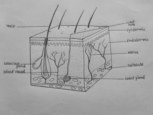
Our skin is the sense organ for touch. It contains cutaneous receptors for touch. The skin consists of two main layers called epidermis and dermis. Epidermis is the layer for protection. It has outer stratum corneum or cornified layer containing dead cells, middle granular layer containing living cells and inner malpighian layer containing the cells dividing constantly. 

Dermis lies below the epidermis. It is made up of elastic connective tissue. It contains sweat glands, sebaceous glands, hair follicles, blood vessels and fat. Here we start the diagram.


1.Draw a cuboid shape as shown.


2.Draw the epidermis and endodermis as shown.


3.Draw hair follicles and other details of the skin
as shown.


4. Label the parts as shown.


How to draw embryo

The growing embryo forms four membranes-Chorion, Amnion, Allantois and Yolk sac.Chorion establishes connection with the walls of the uterus and helps in the supply of nutrients to the embryo and in the removal of wastes from the embryo. Amnion forms a sac like structure around the embryo. The space between the amnion and embryo is filled with a fluid called Amniotic fluid. Amnion and amniotic fluid give protection to the embryo against mechanical shocks. Amnion is retained till the child is born as the embryo grows the amnion also grows in size. Another sac like structure called Allantois is continuous with urinary bladder of growing embryo. Yolk sac usually supposed to contain reserve food material, but in human embryo it is vestigial and useless.


1.Draw tiny human embryo as shown.


2.Make Amnion around it as shown.


3.Draw Yolk sac and Allantois as shown.


4.Draw one more membrane around as shown, it is Chorion


5.Draw tiny projections called chorionic villi as shown and label the parts


How to draw reflex arc

Reflex arc consists of a receptor, a sensory nerve, an association neuron, motor nerve and effector organ. The receptor may be a single cell or a group of cells. They receive information about stimuli and generate electrical potentials. The sensory nerve is associated with receptor. It carries the information in the form of electrical impulses from the receptor organ to the spinal cord. In the spinal cord, the sensory nerve makes connections with other neurons in the grey mater. These neurons are called inter neurons or association neurons. The neurons process the information and generate responses. The efferent fibers of motor nerves carry these responses from inter neurons to the effector organ which is usually a muscle. This makes the muscle to contract.


1.Draw a apple shape as shown.


2.Draw the two tube like shapes as shown on both sides.


3. Position the Receptor and Effector as shown.


4.Connect receptor and effector with nerves as shown.


5. Label the parts as shown.

How to draw TS of spinal cord

Transverse section of spinal cord, shows butterfly shaped or  'H' shaped grey mater. Each segment of H shaped grey mater is called Horn. The upper horns are called Dorsal horns, and the lower horns are called Ventral horns. In the middle of the grey mater is a canal called Spinal canal. It runs all along the length of the spinal cord and is filled with cerebro spinal fluid. 

   From the sides of spinal cord, 31 pairs of nerves take their origin one from each side and supply branches to various parts of the body. Basic function of spinal cord is to act as a relay station-receiving information from various parts of the body parts below the head and send this information to brain. Similarly, it receives information from brain and sends this information to other parts of the body. In addition to this spinal cord also plays a major role in the reflex actions.


1.Draw a inverted apple shape as shown in figure.


2.Make a loop at bottom, make two protrusions at the tip.


3.Draw spinal canal at center and mark four lines in position for dorsal horns.


4.Draw grey mater as shown in figure.


5.Draw ridges along the grey mater as shown.


6. Draw the star patterns in grey matter as shown.


7.Make several curves along contour 


8.Put several neat dots in white mater.


9. Draw outer Pia mater as shown.


10.Label the parts as shown.

The same shape of  TS of spinal cord can be used for drawing  reflex arc.


How to draw animal cell

Animal cells are generally small in size and cell wall is absent. Except the protozoan Euglena no animal cell possesses plastids. Vacuoles in animal cells are many and small. Animal cells have a single highly complex and prominent Golgi apparatus. Let`s draw a typical animal cell. Here we go.


1. Draw a out line of animal cell, put lot of bends as shown to represent flexible  plasma membrane.


2.Draw details of nucleus as shown in figure.


3.Draw Cell organelles as shown.


4. Put lot of neat dots to represent cytoplasm.


5. Label the parts neatly as shown.


How to draw Plant cell

Plant cells are covered by cell wall, it is a unique feature observed in plant cells. While the cell membrane acts as the outer layer in animal cells. The plant`s cell wall is made up of Cellulose. This is considered as major difference between plant cell and an animal cell. The green parts of plants such as leaves, have cells which contain chloroplasts. Which is considered as another unique feature of plant cells. Let`s see how to draw a typical plant cell.


1. Draw a Hexagon as shown in figure.


2. Draw 6 radiating lines  as shown.

3.Draw the double line as shown. Observe carefully the pattern of lines drawn, it may be little confusing first.


4. Make the middle lamellum thick


5. Draw along the inner side of plant cell as shown.


6. Draw another line in side to represent plasma membrane.


7.Draw Nucleus and it's components as shown.
8. Draw cell organelles namely Endoplasmic reticulum, Golgi apparatus, Chloroplasts, Mitochondria, vacuole.
9.Erase cell wall in some places and draw Plasmodesmata that connects adjacent cells.


10.Label the parts as shown.

Share this tutorial with your friends Now!


Matured plant cells of Xylem accumulate Subarin, Lignin cell wall materials and eventually grow more tougher and stronger giving mechanical strength to tree trunks. 


How to draw Nephron


Each kidney is made of about one million single units called nephrons. These are the structural functional units of the kidney and concerned with separating urine from the blood. Each nephron is made of two main parts called malpighian body and covoluted tubule.Malphigian body is double layered cup also called bowman`s capsule. The inner cup consists network of capillaries called Glomerulus.


1.Draw a egg shape and  make a folded curve as shown in the figure and 
   attach it to a vertical line.

2. Draw double line along the curve and draw Glomerulus as shown in the 
    bowman`s capsule.


3. Draw the capillary network as shown in the figure. You may need some patience to represent the intricate network. You can make it simple by drawing the whole capillaries with single thick line, using blunt pencil tip.

4. Label the parts as shown.Read more about nephron.


How to draw LS of kidney


The LS of kidney shows outer dark zone called the cortex and inner pale red zone called medulla which forms the main mass of the kidney. The medulla is made of number of pyramidal structures containing renal tubules or Nephrons projecting into the cavity towards the inner region of kidney called pelvis.This
is the region where renal artery and renal vein enter the kidney. Free end of pelvis shows cup like depressions called calyces. Renal pyramids of medulla project into these calyces.Now, lets`s start drawing the diagram. Here we go.
 

1.Draw a bean shape as shown in figure

2.Erase the depression and make it double lined.
 
3Draw the Pelvis and ureter coming out of the opening.

4.Mark about seven radiating lines along  the inner  curvature.


5.Draw the renal pyramids as shown along the inner curvature of kidney.

6.Draw Calyces connecting the renal pyramids as shown in figure.


7.Mark several lines that represent Nephrons in each renal pyramid and draw 
    renal vein and real artery  as shown.

8.Label the parts neatly as shown in figure below.


How to draw germination of pollen

Pollen grains received by the stigma germinate and give rise to pollen tubes. Only one pollen tube finally reaches the embryo sac. This tube will have two male nuclei, which migrate to the tip of the pollen tube at the time of fertilisation. Usually pollen tube enters the ovule through micropyle. The tube discharges the two male gametes into  its embryo sac. Let`s start drawing the process. Here we go.


1. Draw a oval shape and make a vertical line as shown in first step.

2. Erase the bottom and draw outer integuments of  ovule.

3.Draw inner integument and embryo-sac as shown.


4. Extend the funicle of ovule as shown, this should be connected to ovary later.

5. Draw the ovary around the ovule and extend to style and stigma as shown.

6. Draw outer covering of ovary and style to represent thickness of ovary.


7.Draw pollen grains on stigma and extend a fine line along the style towards
the micropyle to represent pollen tube like a maze puzzle.

8.Draw another parallel curve along the original curve as shown and make two dots
at the tip of pollen tube to represent two male gametes.

9. Label the parts neatly as shown.


Share it Now!